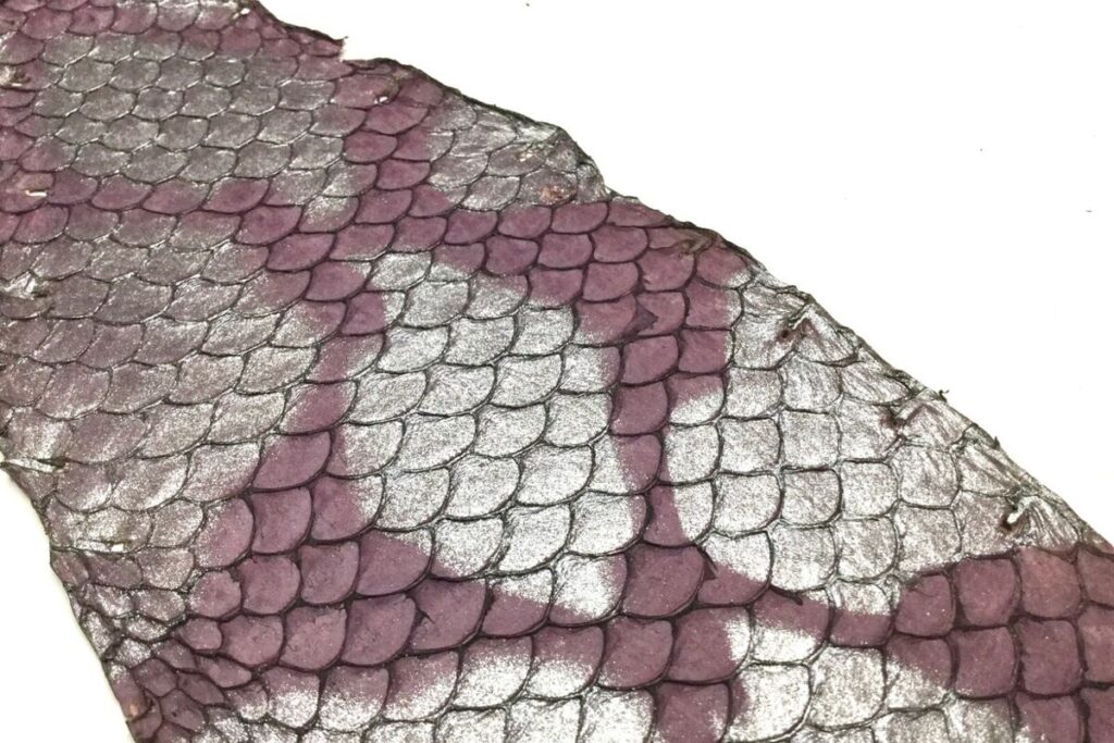
Tilapia skin is rich in collagen and the abundance of this structural protein makes this fish a popular source in veterinary medicine and human medicine. Researchers have found various uses for the skin of this fish, such as dressing burn victims, repairing abdominal hernias, repairing heart valves, and reconstructing vaginas. have discovered.
Mirza Melo, an animal ophthalmologist from Brazil, inspired by his colleagues in dozens of other specialties, tested the effectiveness of tilapia skin in the treatment of corneal ulcers and perforations, especially in dogs with short snouts, a pervasive problem in his specialty. According to him, this group of dogs are animals with big eyes; For this reason, their eyes are often injured.
Corneal injuries caused by external injuries are usually treated by placing a membrane made of horse placenta over the affected area. Horse placenta is a rich source of collagen; But the amount of collagen in it is less than tilapia skin. For the first time in 2019, Dr. Melo operated on a severe corneal hole in a Shih Tzu dog by replacing this membrane with tilapia skin.
After performing his first surgery with the help of tilapia skin, Mello received a call from the Brazilian Burn Support Institute and the Federal University of Ceará (a pioneer in the use of skin in burn treatment). This call, which was made in order to know the technique used in surgery, led to the start of an experiment with the support of this institute. Mello in this cooperation membrane experiment named Cellular dermal matrix (ADM) which was made from pure collagen extracted from fish skin.
According to Mello, collagen is known for stimulating cell growth and directing the production of various tissues. He says that the collagen quality of tilapia fish skin is at a high level throughout the life of this animal. Meanwhile, the quality of horse placenta collagen changes depending on factors such as the age and weight of the animal.
Processed ADM is like a thick sheet of paper that vets soak with saline before surgery and suture after placing it over a dog’s corneal lesion. This cellular dermal matrix acts as a scaffold for cell regeneration. So far, Melo has treated more than 400 dogs with this method, none of which have experienced complications such as pain or infection.
According to Robson Santos, an animal ophthalmologist, current strategies for corneal repair, such as the use of horse placenta and organ transplantation and donation, all have good results; But the scar is still one of the important concerns in these methods. Santos believes that tilapia skin is an excellent alternative to existing methods.
Now, Mello plans to test processed ADM on cats, and says discussions have begun on how to adapt the method to humans. At the same time, he hopes to extend his research to the retina, which is difficult to treat due to the presence of highly sensitive specialized neurons.
According to Mello, the treatment of retinal injuries is where the most limited resources exist in veterinary and human medicine; For this reason, he hopes that one day the use of ADM will become widespread.